Selected Topics

Perception and Meta-Perception

bild

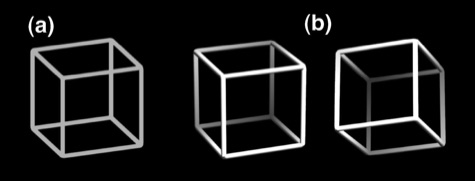
Figure 3. (a) Ambiguous Necker cube and (b) disambiguated cube variants.

During prolonged observation of the ambiguous Necker cube (Fig. 3a, Necker 1832), one of the most prominent ambiguous figures, our perception becomes unstable and reverses spontaneously between two possible interpretations (Fig. 3b). Adding tiny depth cues disambiguates the Necker cube and stabilizes our perception. In a series of EEG studies with very different ambiguous stimuli and disambiguated stimulus variants we studied differences between unstable and stable neural representations (Kornmeier & Bach 2009, Kornmeier et al. 2016, Joos 2020a and Joos 2020b). To our great surprise we a found clear EEG-pattern with small amplitudes of two event-related potentials (ERPs, P200 and P400) in the case of ambiguous stimuli and large amplitudes in the case of disambiguated stimulus variants (Fig. 4, columns 1 – 3).

However, stimulus ambiguity seems not to be the decisive factor, because we found in the meanwhile very similar results using stimuli with varying visibility (e.g. the mouth curvature of happy and sad smiley faces (Fig. 4, left column) or stimuli embedded in noise.

bild
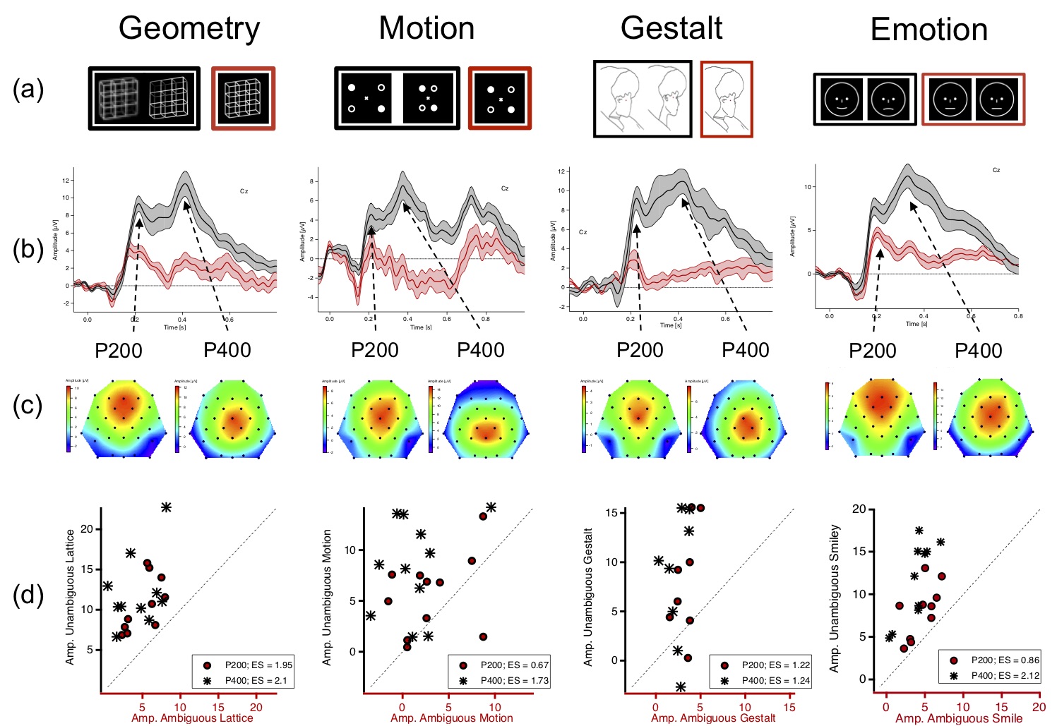
Figure 4. (a) From left to right: Necker lattice (geometry), schematic presentation of the so-called motion quartett (Motion, Von Schiller 1933; online Animation), Boring's Old/Young Woman (Gestalt, Boring 1930), Smiley stimuli. Black/red framed stimuli represent ambiguous/disambiguated stimulus variants. (b) Grand Mean ERP traces (central traces) ± SEM (above and below traces) from EEG elektrode Cz evoked by ambiguous (red traces) or disambiguated (black traces) stimulus variants. The disambiguated stimulus variants evoked significantly larger ERP amplitudes. (c) Voltage maps for the P200 and P400 show in pseudo-colors the distributions of the two ERP components at certain time points (left/right: 200 and 400 ms after stimulus onset). Red/blue colors indicate large/small amplitudes. Remarkable is the similarity of the ERP results across very different stimulus categories (columns). (d) Scatterplots representing individual amplitude values of the different participants (circles/stars represent P200 and P400 amplitudes). The large amplitude differences are also clearly visible on the level of individual participants.

In cooperation with the Department of Psychiatry and Psychotherapy at the Medical Center of the University of Freiburg and the Psychiatric Hospital Strasbourg, France we investigate in this context patients with psychiatric disorders, who also show altered perceptual and conscious states, in order to test our hypotheses and the underlying models. Concurrently, we also try to better understand the focused disorders.

Referenzen

Boring EG (1930). A new ambiguous figure. Am J Psychol. 42, 444–445.

Joos E, Giersch A, Hecker L, Schipp J, Tebartz van Elst L & Kornmeier J (2020a). Large EEG amplitude effects are highly similar across Necker cube, smiley, and abstract stimuli. PLoS ONE 15(5): e0232928.

Joos E, Giersch A, Bhatia K, Heinrich SP, Tebartz van Elst L, Kornmeier J (2020b) Using the perceptual past to predict the perceptual future influences the perceived present – a novel ERP paradigm PLoS ONE 15(9): e0237663.

Kornmeier J & Bach M (2009). Object perception: when our brain is impressed but we do not notice it. Journal of Vision, 9(1),7 1-10.

Kornmeier J, Wörner R & Bach M (2016). Can I trust in what I see? – EEG Evidencefor a Cognitive Evaluation of Perceptual Constructs. Psychophysiology, 53, 1507–1523.

Necker LA (1832). Observations on some remarkable optical phaenomena seen in Switzer-land; and on an optical phaenomenon which occurs on viewing a figure of a crystal or geo-metrical solid. The London and Edinburgh Philosophical Magazine and Journal of Science, 1(5), 329–337.

Schiller PV (1933). Stroboskopische Alternativversuche. Psychologische Forschung, 17, 179–214.


Perceptual Instability and the Generalized Quantum Theory

bild

Figure 5. Grand mean data (± SEM) to indicate the negative correlation between reversal rate during observation of the Necker cube (psychophysical measure) and the latency of a event-related potential (ERP) component (CPP: centro-parietal positivity). Interestingly, this negative relation between measures is predicted on a quantitative level by the Necker-Zeno Model. Red/blue icons = medians of experienced /unexperienced meditators. Light colors: passive observation of the Necker cube; dark colors: participants try to volitionally prevent perceptual reversals (Figure from Kornmeier et al. 2017).

Despite the hithero success story of research in natural sciences and particularly in neuroscience, most fundamental questions concerning consciousness, free will and the psychophysical relations are still unanswered. Interesting in this context is the common theoretical work by the physicist and nobel prize laureate Wolfgang Pauli and the famous psychiatrist Carl Gustav Jung (e.g. Atmanspacher & Fuchs 2014). Atmanspacher and colleagues advanced these ideas and formulated the Generalized Quantum Theory (“GQT”, Atmanspacher et al. 2002). The Generalized Quantum Theory is a formal framework, that allows the application of mathematical concepts also beyond physics and that resulted in empirically testable hypotheses. A very good review article from Atmanspacher can be found in the Stanford Encyclopedia of Philosophy (Atmanspacher 2020). Atmanspacher et al. used perceptual bistability of ambiguous figures as model situation for their theory and developed the so-called Necker-Zeno Model of Bistable Perception, that is based on the GQT. The model presents a relation between observables from three different time scales, that are frequently discussed in Psychology and Cognitive Sciences. In recent years we were able to confirm the model both with psychophysical but also with EEG data. Particularly the authors of the Zeno Model predicted higher values at certain time scales in experienced meditators, which has been also confirmed in our recent study (Kornmeier et al. 2017).


Referenzen

Atmanspacher, H (2020). Quantum Approaches to Consciousness. The Stanford Encyclopedia of Philosophy.

Atmanspacher H, Bach M, Filk T, Kornmeier J,& Römer H (2008). Cognitive time scales in a Necker-Zeno Model for bistable perception. The Open Cybernetics and Systemics Journal, 2, 234–251.

Atmanspacher H& Fuchs CA (2014). The Pauli-Jung conjecture and its impact today. Imprint Academic.

Atmanspacher H, Römer H & Walach H (2002). Weak quantum theory: Complementarity and entanglement in physics and beyond. Found Phys, 32, 379–406.

Kornmeier J, Friedel E, Wittmann M & Atmanspacher H (2017). EEG correlates of cognitive time scales in the Necker-Zeno model for bistable perception. Consciousness and Cognition 53, 136 - 150.

Data Analysis with Methods from Artificial Intelligence

bild

Figure 6. Example of a feed-forward fully connected neural network (from Hramov et al. 2019). Blue traces (left) are example EEG traces from one participant, si, ui and y are neurons of the artificial network. ωi represents the weighting für the information transfer between neurons of successive network layers. During training of an artificial neural network the ωi will be stepwise altered to improve the network performance.

Scientific data (in our case EEG or behavioural data) contain patterns, that may be invisible for our eyes. Sophisticated analysis methods allow to detect such patterns and sophisticatd graphical methods allow their visualization. The analysis results typically depend strongly on the method of choice and the related restrictions and pre-assumptions. Methods from artificial intelligence (AI) – and particularly from machine learning (ML) – show an increasing power for data analysis in various fields. ML-methods are also more and more often used in neuroscience and particularly in clinical studies, e.g. for the segmentation of 3D tomographic images or for classification purposes (Milletari et al. 2016, Qureshi et al. 2019, Rad & Furlanello 2016).

However, the larger power of ML-solutions compared to more classic approaches comes together with long-lasting sessions to train the artificial neural networks. High-performance computers and graphic boards become more and more important in this context. In autumn 2019 we successfully applied for an Eucor–Seeding Projekt , to apply ML-solutions to clinical data. We also received a high-performance graphics board (Nvidia Titan V) during a call from the NVIDIA company. We currently use this graphics board to analyse given EEG data with ML-methods and further developed an artificial network to calculate brain sources for EEG signals.

Referenzen

Milletari F, Navab N & Ahmadi SA (2016). V-Net: Fully Convolutional Neural Networks for Volumetric Medical Image Segmentation. 2016 Fourth International Conference on 3D Vision (3DV), 565–571.

Rad NM &Furlanello C (2016). Applying Deep Learning to Stereotypical Motor Movement Detection in Autism Spectrum Disorders. 2016 IEEE 16th International Conference on Data Mining Workshops (ICDMW), 1235–1242.

Qureshi MNI, Oh J & Lee B (2019). 3D-CNN based discrimination of schizophrenia using resting-state fMRI. Artificial Intelligence in Medicine, 98, 10–17.

ConvDip: A Convolutional Neural Network to better analyze EEG Sources

Patterns in EEG data raise questions about underlying functions and brain sources. Which brain area is the source of a signal measured on the scalp? This question, also know as the inverse problem of EEG, is discussed by scientists since the beginning of EEG recording. The problem as such is ill defined because one and the same EEG pattern on the scalp can theoretically be evoked by the concurrent activity two or more different configurations of brain sources. A unique solution does not exist. Several approaches to solve this inverse problem are at hand, each with advantages and disadvantages. One problem of an often-used approach (the Minimum Norm Solution) is the broad spatial distribution of the reconstructed sources, which is not confirmed by findings and theoretical models of neural signals (e.g. Nunez & Srinivasan, 2006).

With “ConvDip” (Hecker et al. 2020) we have developed a method to solve the inverse problem with a convolutional neural network. We construced physiologically plausible artificial sources and calculated the corresponding EEG signals for the EEG electrodes on a theoretical scalp (forward solution). With these data we trained our network. With a separate set of data we then tested the performance of the trained ConvDip network and compared the performance of ConvDip with the performance of other classical source localization methods. ConvDip outperformed the classical methods for all used measures (see Fig. 7 for examples).

bild
Figure 7. Left: Boxplots of the variances that are explained by the projections of the found sources on the scalp for each of the tested source analysis methods. The horizontal line (at 81%) marks the signal-to-noise ratio in the artificially generated EEG data. ConvDip explains significantly more variance than the other two methods, independent of the number of modeled sources. Right: normalized average squared error for the different inverse solutions. ConvDip produces significantly less normalized averaged squared errors than the other methods. The white horizontal lines in the box plots indicate the medians, the rectangles above and below the inter-quartile ranges. The whiskers indicate the central 95% of the data.

References

Hecker L, Rupprecht R, Tebartz van Elst L, Kornmeier J (2021) ConvDip: A convolutional neural network for improved M/EEG Source Imaging. Frontiers in Neuroscience 15.

Hramov AE, Maksimenko V, Koronovskii A, Runnova AE, Zhuravlev M, Pisarchik AN & Kurths J (2019). Percept-related EEG classification using machine learning approach and fea-tures of functional brain connectivity. Chaos: An Interdisciplinary Journal of Nonlinear Science, 29(9), 093110.

Nunez P & Srinivasan R (2006). Electric Fields of the Brain: The Neurophysics of EEG. Ox-ford 561, University Press, USA, 2006.

Milletari F, Navab N & Ahmadi SA (2016). V-Net: Fully Convolutional Neural Networks for Volumetric Medical Image Segmentation. 2016 Fourth International Conference on 3D Vision (3DV), 565–571.

Qureshi MNI, Oh J & Lee B (2019). 3D-CNN based discrimination of schizophrenia using resting-state fMRI. Artificial Intelligence in Medicine, 98, 10–17.

Rad NM &Furlanello C (2016). Applying Deep Learning to Stereotypical Motor Movement Detection in Autism Spectrum Disorders. 2016 IEEE 16th International Conference on Data Mining Workshops (ICDMW), 1235–1242..